|

Tibial Plateau Leveling Osteotomy (TPLO) Surgery (*The most common orthopedic surgery at our clinic)
 The TPLO is my preference for managing a patient with a torn anterial/cranial cruciate ligament(ACL/CCL). However some cases I perform TTA (Tibial Tuberosity Advancement) surgery over TPLO depending on shape of the joint and angles of the joints and especially a knee has ACL and patella luxation (MPL/LPL) at the same time. This is based on my experiences of performing a variety of ACL repair techniques for over the years. I currently performs many TPLOs ranging in pet size from 12-140 pounds. The TPLO is my preference for managing a patient with a torn anterial/cranial cruciate ligament(ACL/CCL). However some cases I perform TTA (Tibial Tuberosity Advancement) surgery over TPLO depending on shape of the joint and angles of the joints and especially a knee has ACL and patella luxation (MPL/LPL) at the same time. This is based on my experiences of performing a variety of ACL repair techniques for over the years. I currently performs many TPLOs ranging in pet size from 12-140 pounds.
In the dog’s knee joint, the surface of the tibia or shin bone, called the tibial plateau, has a slope, called the tibial plateau angle (TPA). The TPA varies in each dog and varies even within knees of the same dog. In most cases, the TPA ranges from 18 degrees to 33 degrees from a line drawn perpendicular to the long axis of the tibia. There are some TPAs that measure greater than this. When a dog weight bears, the tibia wants to thrust forward on this slope. The CrCL normally restrains this from happening. The principle behind a TPLO surgery is to cut the tibia bone with a curved cut and rotate it to level the slope thus preventing the shifting of the bones. The rotated bone is secured into its new position with a bone plate and screws while the bone heals. The changing of the geometry of the bone alters the biomechanical forces acting around the knee joint. The TPLO levels the slope so that the femur no longer slides down the plateau or the tibia is not thrusting forward in relationship to the femur. Thus a dynamically stable joint is created even with the absence of a CrCL. Consequently, we do not rely on the CrCL anymore. The change in the mechanical forces allows for the stability in the knee.
WHAT HAPPENS DURING THE SURGICAL PROCEDURE?
 Dr. Song performs the surgery with assistance from his trained surgical technician. Dr. Song examines the health of the cartilage in the joint, debrides the torn ligament, and assesses the health and integrity of the menisci in the joint. Approximately 50% of the patients have torn medial menisci and the damaged portion is removed. Following this, the TPLO procedure ensues. The surgery time itself takes approximately 2 hours and total anesthesia from induction through preparation and radiographs lasts approximately 2-2 1/2 hours. Dr. Song performs the surgery with assistance from his trained surgical technician. Dr. Song examines the health of the cartilage in the joint, debrides the torn ligament, and assesses the health and integrity of the menisci in the joint. Approximately 50% of the patients have torn medial menisci and the damaged portion is removed. Following this, the TPLO procedure ensues. The surgery time itself takes approximately 2 hours and total anesthesia from induction through preparation and radiographs lasts approximately 2-2 1/2 hours.
AFTER SURGERY WHAT IS THE AFTERCARE REQUIRED ONCE WE BRING OUR PET HOME FROM THE HOSPITAL?
Most of the time, your pet will stay one night in the hospital or overnight care facility.
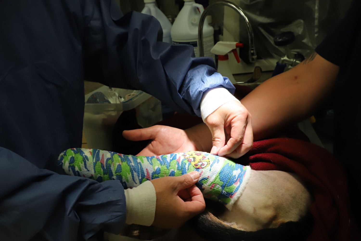
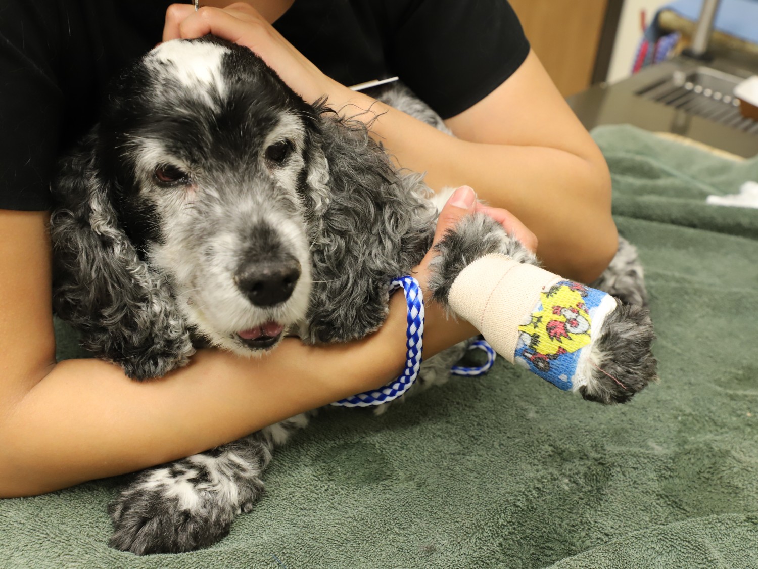
Your pet will have a bandage covering the wound. This should be removed within 3-4 days.
Your pet will go home with a course of antibiotics, an anti-inflammatory drug, and some oral pain medication. Sometimes additional methods of pain control are prescribed.
Do not allow licking at the incision, this increases the risks of infection.
It is not unusual to see a little swelling in the lower hock or ankle joint starting at 3-5 days after surgery. This is migrating fluid or edema from the surgery site. Warm moist compresses will enhance increased blood and lymphatic flow to assist in resolving this within a couple days.
There will be staples or stitches in the wounds at the site of the main surgery.
You will recheck with your Veterinarian at 12-14 days to have the staples or stitches removed.
You will recheck at 8 weeks from surgery to have radiographs taken of the surgery site to assess bone healing.
If all is progressing well, a recheck at 10 and 14 weeks may be required before the patient can return to normal activity.
WHAT LEVEL OF ACTIVITY RESTRICTION IS REQUIRED WITH A TPLO?
Think of the aftercare as Two Phases.
Phase I: Allowing the bone to heal: (First 6 Weeks)
(See an example Discharge Instruction sheet at the bottom of this manuscript)
The focus during the first phase is to create an environment for the bone to heal adequately. Therefore; no running, jumping, rough play, multiple stairs, or off leash activity when your pet is outside is expected. Walking around the house is acceptable, however, when unsupervised; consider crate or small room confinement. Your pet should always be on a leash when outside and outside only to eliminate and carefully walk around on the leash for 5-10 minutes.
Phase II: Strengthen and Condition the soft tissues: (6-16 Weeks)
After the bone has an early union and of adequate strength, the focus is to properly condition and strengthen the soft tissues of the leg and the knee which are experiencing different mechanical forces now that the geometry of the bone has changed. At this stage, once radiographs reveal the presence of adequate early bone healing, progressively longer leash walks are allowed. This is when more involved physical therapy can begin as well. Still no off leash activity during this time is expected. Patients can return to normal activity in the 14-16 week time line depending on their progress.
CAN WE WORK WITH A PHYSICAL THERAPIST AFTER SURGERY?
Yes, you may consult with a Physical Therapist. Physical Therapists add a valuable service to the recovery of our patients, although not required, there is strong supportive evidence that physical therapy will improve upon and shorten the rehabilitation process and help our patients transition to normal activity by improving on muscle mass and strength and to prevent reinjury once return to normal activity is allowed. However, during the first 4-6 weeks while the bone is healing, we do not want to over stress or create unnecessary forces that could jeopardize bone healing, nor do we want to push too hard as this can cause setbacks with strain in the muscles and sprain in the patellar ligament.
During the first 4-6 weeks, passive range of motion or flexion and extension of the knee can be performed. These should be quality, lower numbers of repetitions. Other activities include single leg stances, lifting non surgical leg, controlled backward walking, etc. After 6 weeks, more activities are introduced in a controlled manner, such as walking, front feet on the box, underwater treadmills, swimming, etc.
MY VETERINARIAN WOULD LIKE TO USE LASER THERAPY?
Laser therapy can decrease inflammation, and enhance the healing of the surgery site. Some hospitals will offer laser therapy treatments and this can begin immediately after surgery and continued for a variable length of time throughout the recovery, depending on how well the patient is progressing. Please consult your Veterinarian and or a Physical Therapist.
WHAT SHOULD WE EXPECT FOR PROGRESS AND USE OF THE LEG AFTER SURGERY?
 Some patients will be placing the leg to the ground at the walk, the day after surgery, others take several days. It would be unusual for a patient to be holding up the leg at the walk by staple or stitch removal, if this should occur, see your Veterinarian and ask them to contact us for discussion and possible re evaluation if necessary. After Staple or stitch removal, gradual increased use of the leg should be noted throughout the 14-16 week process. By 16 weeks when full uncontrolled activity is allow, the patient should experience a good outcome with limited signs of lameness in 90%+ of the patients. We have seen hunting dogs successfully hunt again. Some patients will be placing the leg to the ground at the walk, the day after surgery, others take several days. It would be unusual for a patient to be holding up the leg at the walk by staple or stitch removal, if this should occur, see your Veterinarian and ask them to contact us for discussion and possible re evaluation if necessary. After Staple or stitch removal, gradual increased use of the leg should be noted throughout the 14-16 week process. By 16 weeks when full uncontrolled activity is allow, the patient should experience a good outcome with limited signs of lameness in 90%+ of the patients. We have seen hunting dogs successfully hunt again.
WHAT ARE THE SUCCESS RATES?
Although all surgeries carry with them a certain level of risk and complication, each patient has a different degree of arthritis present at the time of surgery, varying levels of activity present in their lifestyle (hunting, hiking, playing, walking, running, etc), different size and weight conditions are present, and other orthopedic or medical problems may coexist. In general, 90%+ of our surgical patients will return to a high level of comfortable athletic activity. In our experiences, the risks of complications are approximately 3-5%. This may include surgical site infection, the need for implant removal, excessive inflammation, menisci injuries, to name a few. It is very uncommon that a second surgery is required based on Dr. Song's experience.
WHAT IS THE LIKELIHOOD THAT THE OTHER KNEE WILL ALSO DEVELOP A RUPTURE OF THE CRANIAL CRUCIATE LIGAMENT AND REQUIRE A TPLO?
Studies have shown that approximately 50% of the patients will tear the opposite cruciate ligament within a year or two timeline from the diagnosis of the original tear. Sometimes our patients have torn ligaments in both knees at the time of our diagnosis. I prefer that we perform surgery on the leg that the patient is most lame or most clinical and then 4-8 weeks later surgery can be performed on the other knee. Surgery can be delayed longer, but 4 weeks is my preference for the shortened interval. On rare occasion, the patient is so debilitated with significant trouble getting up and walking that we will perform the surgery on both knees at the same surgery. This may add risk and is not ideal, however; we have performed surgery bilaterally with equal success in many cases.
WILL THE PLATE REMAIN IN MY PET?
Yes, we do not remove the plates unless problems arise which dictate their removal. Although infection rates are low, implants may harbor bacteria that are not easily remedied without plate removal. We can see infections develop months to years after the surgery, not just postoperatively. This is likely due to the fact that bacteria may circulate in the blood system and set up at the site of the implant. When teeth are cleaned, when patients chew food, when bowel movements occur, urinary tract infections, or skin infections occur, all these can add risk. The bacteria secrete a protective biological layer that prevents the ease for white blood cells and antibiotics to penetrate. Removal of the plate allows for improved access. We remove only 1-2% of plates or 3-4 out of 250 cases. Plate removal can be performed with high success and low complications.
Although there is anxiety amongst a few that implants can increase the risk of developing bone cancer, there is no scientifically proven data that this is the case. Reports have not proven the cause and effect. Anytime tissue is healing there is inflammation and there is a risk that cells can undergo change and become cancerous. This is extremely rare. We have seen cancers develop from vaccine injections, broken bones that had no surgery and were placed in a cast for example, only to see bone cancer developed later and no implant was associated. We do not downplay the risks, but they are so small considering the tens of thousands of plates placed each year, that we must balance this against the risks of empirically removing all plates in all of our patients. I have been practicing since 1985 and I have not seen this to be a problem. I want my clients to be educated, but not to be overly worried.
HERE IS A COPY OF THE STANDARD REPORT THAT WILL BE SENT HOME WITH YOU:
TPLO DISCHARGE INSTRUCTIONS
A Tibial Plateau Leveling Osteotomy (TPLO) has been performed to correct the instability of your pet’s knee. This surgery entails a curved bone cut through the top of the tibia, which allows the joint surface to be rotated into a more level position. The bone is then stabilized in its new position with an orthopedic plate and 6 screws. It will take your pet approximately 8 weeks to achieve early bone healing and approximately 16 weeks to achieve mature bone healing. The patient may be placing the foot to the ground at the walk within a few to several days after surgery.
There are several aspects of your pet’s recovery that are imperative:
PHASE I: Allowing the Bone to Heal!
1. Do not allow your pet to lick or chew at its incision until the skin has healed (14 days). Skin staples or stitches may be removed in about 10-14 days after surgery. Some swelling of the hock is normal between days 3 and 7 post-operatively. It should begin to resolve approximately 10 days after surgery. However, if you notice excessive redness or any discharge from the incision, your pet should be re-evaluated by your regular veterinarian.
2. **Do not allow your pet to have any vigorous activity for 6-8 weeks (Based on x-ray evaluation).
- Outside only on a leash; only long enough for eliminations, never off leash!
- Confine to a small area (large dog crate) when not directly supervised.
- Some freedom indoors is permitted under your supervision – no using stairs, no jumping on furniture, be careful on slippery surfaces, no running or rough play.
3. Recheck X-rays should be taken at 6-8 weeks after surgery or earlier if we have any concerns. Sometimes X-rays are taken at 4 and 8 weeks, but a single X-ray at 8 weeks may be adequate if the patient is doing well with weight bearing and good progress is being made with bone healing.
PHASE II: Strengthen and Condition the soft tissues, primarily the Quadriceps.
4. After evaluation of the 8 week X-rays, you may begin leash walking your pet. (At this time frame, you may consider consultation with a Physical Therapist also. References are available)
- Begin with a short distance for 3-5 days. If no lameness is encountered, the distance may be increased.
- Every time the distance is increased, monitor your pet’s response for 3-5 days before increasing the distance again. Controlled use of stairs, if necessary, is okay now.
- Continue strict confinement when not supervised.
- Gradually increase back to normal activity up until 16 weeks.
- Off leash activity may begin 16 weeks after surgery.
Summary
1. Surgery Date
2. Staple/Stitch Removal: ~ 10-14 days
3. Recheck X-rays 8 weeks as long as good progress is being made and no concerns or problems exist otherwise at 4 and 8 weeks they should be taken.
4. Strict confinement until it is determined that the X-rays show early bone healing.
Leash walking until 16 weeks at which time off leash activity is allowed. The basis for the continued controlled leash walk activity is to build strength in the ligaments and muscles which are experiencing different mechanical stresses from the surgical procedure and to allow for mature bone healing.
|