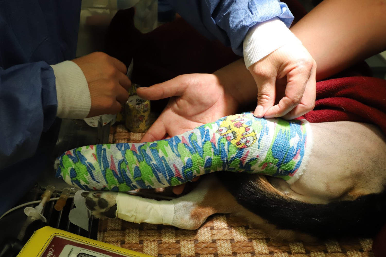
Hind Limb Lameness
One of the most common causes of hind limb lameness in the dog is Anterial/Cranial Cruciate Ligament (ACL/CCL) rupture. The CCL, the equivalent of the anterior cruciate ligament (ACL) in humans, and the Caudal Cruciate Ligament (CaCL) sit inside the stifle (knee) joint and prevent the proximal tibia from sliding back and forth with respect to the distal femur. At the top of the tibia is the tibial plateau, a condylar, cartilage covered surface that supports the weight coming from the femur. The plateau is convex as are the condyles of the femur and in weight bearing, the tendency of the femur is to slide down the tibial plateau pushing the top of the tibia forward (cranial tibial translation). The CCL stops this downward slide, occurring in locomotion for millions of cycles per year. This repetitive biomechanical stress on the ligament often leads to fraying and eventual tear or rupture of the CCL. Rupture is a major cause of Degenerative Joint Disease (DJD).
Signs of CCL Rupture:
- Sudden lameness on one rear limb to a degree that weight can not be borne.
- A history of mild lameness in the same limb that seems to come and go before this sudden worsening of symptoms.
Consequences of CCL Rupture:
- Ligament deterioration releases a combination of inflammatory factors from the ligament.
- Increasing instability and inflammation of the joint from the weakened ligament causes arthritis to develop quickly within the joint.
- Every time the pet bears weight on the affected leg, the femur slides down the tibial plateau with nothing to halt its movement. This sliding action damages a cartilage seal in the joint called the meniscus. Once the meniscus is torn, arthritic change accelerates and perceived pain worsens.
- Weight bearing studies, using force plate testing, have shown that dogs with a damaged CCL bear only 20-30% of the normal amount of weight on the affected leg. As a result, the weight is shifted to the other rear leg placing even more stress on that limb's CCL and increasing the possibility of damage to that stifle. Rupture of the CCL in both stifles is common.
Treatment Options:
Veterinarians have been using many treatment options to minimize deterioration of the stifle in dogs with CCL damage. These range from very conservative treatment, prescribing pain management drugs, to aggressive surgical treatment, cutting bone to alter joint biomechanics, with other less invasive surgical procedures in between. Although pain management drugs may help the dog feel better and cope with a bad knee, they do not alter the progression of disease.
Many surgical suture techniques have been developed. Newer procedures use very strong braided materials to hold the tibia in place relative to the femur. Suture techniques may have positive short term outcomes followed by the development of joint fibrosis that can increase stability, but progression of arthrosis is commonly observed, especially in young and active medium or larger dogs.
The first, broadly accepted, orthopedic procedure to correct knee geometry was the Tibial Plateau Leveling Osteotomy (TPLO) of Dr. Barclay Slocum. A curved cut, bisecting the proximal end (top) of the tibia, separates the tibial plateau from the rest of the bone. The plateau is rotated, removing the natural slope of the plateau. A custom shaped metal plate is used to secure the plateau segment back to the tibia with screws.
TPLO has been a successful technique with about 30 years of history. It is a complex procedure that can lead to major complications if not performed correctly.
For a detailed comparison of TTA and TPLO see>> Boudrieau RJ, - Tibial Plateau Leveling Osteotomy or Tibial Tuberosity Advancement? Vet Surg 38:1-22, 2009.
Other corrective procedures include Closing Wedge Osteotomy (CWO), Triple Tibial Osteotomy (TTO), Tibial Wedge Osteotomy (TWO), and Fibular Head Transposition.
***All copyrights are reserved to Kyon International Inc.


